Search Results
See All Results
Home
Reels
Groups
Pages
See More
Groups
Pages
Events
Blogs
Funding
Offers
Jobs
Forums
Movies
Join
Sign In
Sign Up
Theme Switcher
Day Mode
Search
Posts
Blogs
Users
Pages
Groups
Events
dry_magician_7086
added a photo
2026-02-22 05:09:03
·
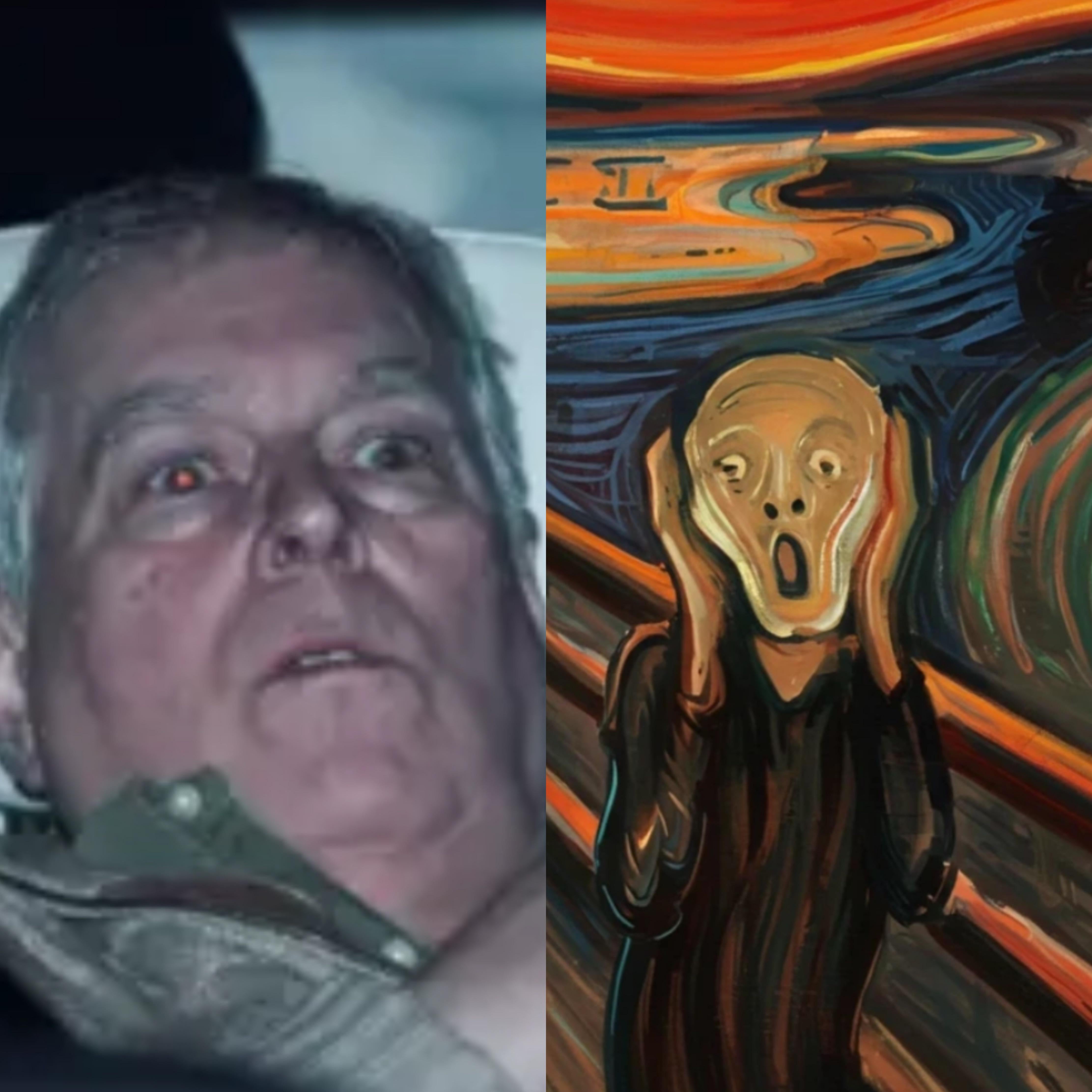
Life imitates art
Life imitates art
3
·
0 Comments
·
0 Shares
·
131K Views
·
0 Reviews
nessakill
added a photo
2026-02-20 14:31:03
·
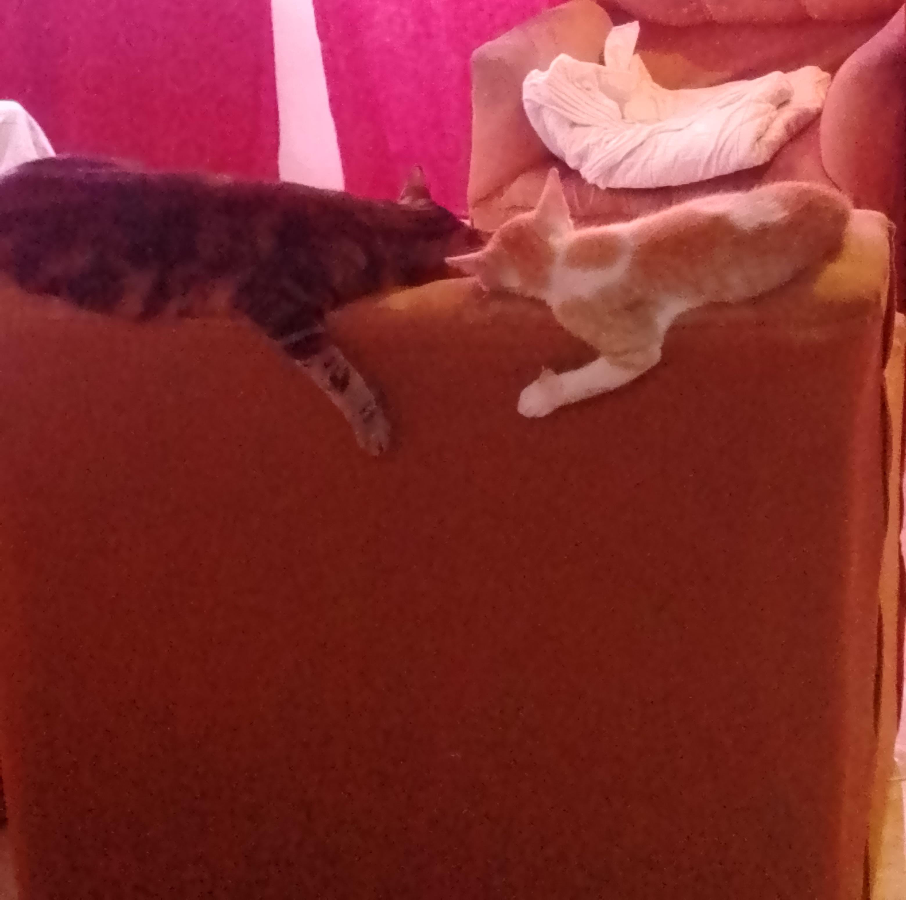
mi eduardo siempre imitando a su hermana mayor
mi eduardo siempre imitando a su hermana mayor
3
·
0 Comments
·
0 Shares
·
130K Views
·
0 Reviews
PoopSoBigCantFlush
added a photo
2026-02-03 16:36:03
·
Damn you, indomitable human spirit!
Damn you, indomitable human spirit!
3
·
0 Comments
·
0 Shares
·
106K Views
·
0 Reviews
cristopher42_b0fx
added a photo
2026-01-22 10:45:03
·
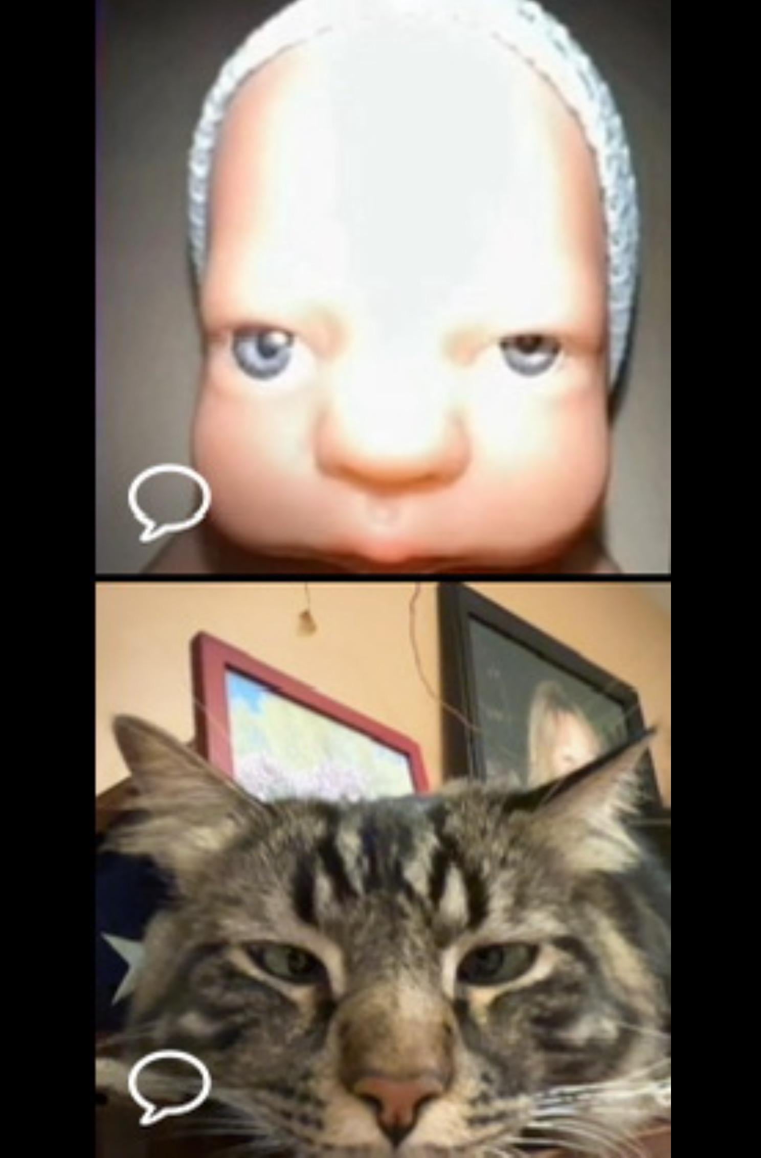
When art imitates my cat
When art imitates my cat
3
·
0 Comments
·
0 Shares
·
113K Views
·
0 Reviews
morris_orn_f6pg
added a photo
2026-01-19 20:56:03
·
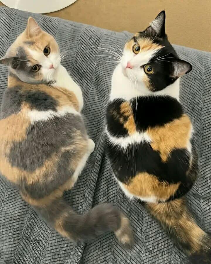
Original and imitation
Original and imitation
3
·
0 Comments
·
0 Shares
·
112K Views
·
0 Reviews
More Results
Upgrade to Pro
Choose the Plan That's Right for You
Upgrade